In biomedical research, the viability and recovery of peripheral blood mononuclear cells (PBMCs) determine the reliability of downstream results. Over the past few years, advances in donor screening, cryopreservation, and logistics have dramatically improved the consistency of primary immune cell performance.
Yet, small variables—temperature shifts, prolonged DMSO exposure, or donor differences—still account for major discrepancies in experimental reproducibility. At CGT Global, we continuously evaluate the factors that most influence PBMC quality to help researchers obtain high-viability, high-recovery results from every sample.
This updated guide explores the latest insights on blood collection, transport, isolation, freezing, and thawing — along with how standardized donor programs and validated protocols ensure reproducible, high-quality outcomes in 2026.
By Tera Muir, PhD./CGT Global
Why is PBMC Viability and Recovery Important?
Blood contains the precious cells which keep us alive. Red blood cells that supply our body with oxygen, our white blood cells that fight invaders, plasma that maintains blood pressure and regulates body temperature, and platelets essential for blood clotting. Taking these critical elements out of their natural environment can lead to a number of issues.
The immediate environment can influence cellular function because of the wide variety of receptors on the exterior of the cell membrane. Receptors allow the cells to communicate with their environment and direct communication with neighboring cells by receiving and processing relevant information, which, as a response, change their internal workings. Therefore, changing the environment of a cell can have a significant effect on the cellular processes of neighboring viable cells. Changes in the cellular processes of viable cells can cause adverse effects on your experiments and assays.
Cell viability can directly impact the recovery of cells. When cells die, their membranes open, allowing the DNA to move out into the extracellular environment. DNA is very sticky and can clump viable cells together, causing a decline in recovery. Too few cells can impact your downstream application.
Blood Collection
Most immune cells circulate within our blood. To collect these cells, whole blood, or an apheresis product, is collected by venipuncture into an appropriate container (bag, tube, etc.) that contains an anticoagulant. The whole blood or apheresis product should immediately mix with the anticoagulant to prevent clotting. Although straightforward, there are issues that can arise and affect cell viability and recovery.
A common issue that can occur when collecting blood is a slow blood draw, which is typically caused by the small vein size of the donor. Since coagulation begins almost immediately, a slow blood draw can interfere with the mixing of the anticoagulant with the blood causing thickening of the blood and blood clots, inevitably leading to poor separation and recovery of cells when trying to isolate PBMCs. There is no immediate remedy to fix a slow draw, but to help prevent clots invert the tube or bag a few times while proceeding with the draw to promote mixing of the anticoagulant.
And don’t forget that the size of the needle can impact the quality of the blood cells. After noticing hemolysis occurring in my blood sample, and hours of pondering what might have gone wrong, a quick phone call to the tube supplier set me straight. Too small of a needle can cause excess vacuum force, while too large of a needle can cause shear stress on the cell walls, both of which lead to hemolysis. A standard 21- or 22-gauge needle for routine collection resolves the issue.
Apheresis is more “high tech” means of isolating PBMCs than a simple whole blood draw. An apheresis machine continuously draws blood out of a donor, spins the blood to separate the white blood cells from the red blood cells and plasma, returns the red blood cells and plasma to the donor, and transfers the white blood cells to a collection bag, often referred to as a Leukopak. A highly trained technician can adjust parameters during the collection procedure to remove more red blood cells and granulocytes, providing a highly pure collection of PBMCs. We don’t see many problems regarding the viability of cells from modern-day apheresis machines. And the only issue with the amount of PBMCs we can collect is because of donor variability.
Blood Transportation & Storage
Consider yourself lucky to receive blood for your research on the same day of collection from a donor. For most, the blood is transported from a collection center to the research facility for further use. Transport of whole blood products, such as bone marrow, peripheral blood, cord blood, and apheresis products, at room temperature (15-25°C) and <24 hours of collection, is the best choice for preserving cell integrity.
For fresh blood, initial gentle mixing with an anticoagulant during collection is important to prevent the blood from clotting. If you plan on storing blood for >24 hours, merely set the blood aside at room temperature after the initial mixing, and it should be ready for use when you need it. It might seem plausible to store the blood on a continuous rocker, however continuous mixing of the sample well after collection can cause micro-clotting. Even slow, gentle rocking over time will cause micro-clotting. The presence of micro-clots can impact downstream experiments and cause cell loss when trying to isolate PBMCs.
For most researchers who do not have direct access to fresh blood often choose overnight delivery. Ambient or room temperature is often preferred. However, if using a courier, transport at room temperature can fluctuate depending on the season and location. Some parts of the country can get well below freezing in winter, while in the summer, temperatures can reach into the 100s. Extreme temperatures can be detrimental to blood cells. Viability and, therefore, recovery will decline. To preserve the viability of the cells when transporting overnight, a 2-8°C or 15-25°C validated shipper is recommended. The validated shipper prevents the exposure of the cargo to extremes in temperature.
However, transporting or storing the blood at 2-8°C for >24 hours can intensify granulocyte contamination in the PBMC fraction. This prolonged storage of whole blood can activate granulocytes, which affects their buoyancy profile resulting in inefficient separation and recovery of the PBMC fraction when using a density gradient.
For Leukopaks, freezing the sample after collection is a plausible option when transporting immune cells over long distances. As mentioned previously, Leukopaks contain minimal amounts of red blood cells and granulocytes. However, when present, cells such as granulocytes can create issues for other immune cells. For example, T cell proliferation following PHA stimulation significantly declines in the PBMC fractions that contain contaminating granulocytes. And there is a correlation between granulocyte contamination and loss of cell integrity, cell number, and reduction of variability in Regulatory T cells. Therefore, freezing can be beneficial when having to transport Leukopaks over a distance that can take up to 24 hours or longer.
Standardizing Donor Quality and Handling
Donor variability remains one of the largest contributors to inconsistent cell recovery and performance. To address this, labs are adopting controlled donor programs and standardized handling methods to reduce variability from the very start.
At CGT Global, verified donor programs and recallable donor pools ensure consistency in age, health, and collection conditions. Every donor sample includes documented collection parameters and traceable quality metrics.
This single-donor consistency, combined with validated processing protocols, helps eliminate variability at the earliest stage—improving reproducibility across downstream studies.
Cell Isolation
Two primary techniques that separate PBMCs from whole peripheral blood are through the use of a density gradient centrifugation procedure or by apheresis. You can find more information about the different techniques HERE.
When working with whole blood, it is necessary to perform a density gradient to enrich for PBMCs. There are different brand names for this type of solution, such as Ficoll and Histopaque, but mainly they work the same way. Each brand (Ficoll and Histopaque) comes with very detailed information regarding troubleshooting tips. I highly recommend reviewing the information before starting any procedure.
The most common issue to affect the density gradient procedure is temperature. Blood, buffers, and other reagents are often stored at 2-8°C, and if not allowed to equilibrate to room temperature (15-25˚C), it will cause a poor separation of the blood. At room temperature, red blood cells aggregate in the presence of a density gradient. Aggregation increases the rate of sedimentation of the red blood cells, which rapidly collects as a pellet at the bottom of the density gradient. These aggregates will not form if cold blood is used, allowing red blood cells to move into and contaminate the PBMC fraction.
The age of the blood is another factor that can influence the outcome of a density gradient procedure. Blood that is drawn <24 hours before separation will provide the best results when performing a density gradient procedure. However, for many researchers, access to freshly drawn blood is not possible. If the drawn blood is >24 hours, the separation will be more difficult and can cause recovery and viability to decline as well as an increase in contaminating granulocytes. To possibly increase viability, cells can be run over a filter to remove any clumps caused by dead cells. Additionally, residual granulocytes can be depleted out of the PBMC fraction by using CD15 or CD16 MicroBeads. Keep in mind that using a filter and/or depleting out granulocytes will cause a decline in recovery.
Leukopaks contain an enriched PBMC fraction, but it can also have some contaminating granulocytes, RBCs, platelets, and plasma. For many applications, an RBC lysis and multiple washes will clear the contaminants. Few granulocytes will be present, roughly 3-10% of total cells. If this is an issue, the use of a density gradient can clear up the contaminating granulocytes. Again, the factors that commonly affect how well a density gradient works for whole blood also applies to a Leukopak.
Freezing Cells
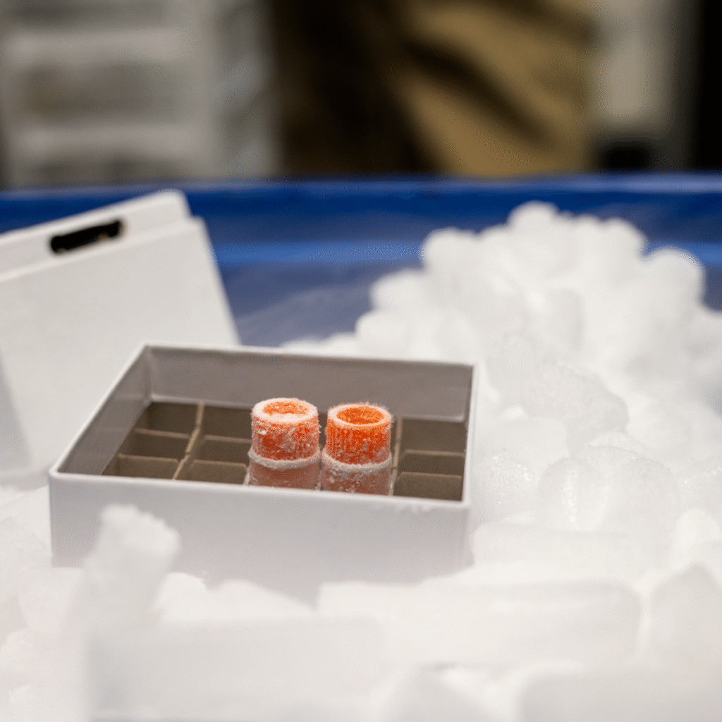
Cryopreservation is the preservation of intact living cells at cryogenic temperatures. Since water is the most abundant molecule in cells, the cells are frozen in the presence of a cryoprotectant which reduces intracellular ice formation and osmotic stress during the freezing process. Cells are then stored at temperatures exceeding -135°C, usually in the vapor phase of liquid nitrogen. This process allows storing the cells for a long duration of time, often for years, in a state of normal metabolic arrest to protect them from damage due to chemical reactivity and time. The cells to be thawed and revived as needed.
Dimethyl sulfoxide, widely known as DMSO, when used at a concentration of less than 10%, becomes a low toxicity cryoprotectant. However, if left for too long in 10% DMSO before freezing, and this means a few minutes for some sensitive cell types, the DMSO becomes toxic, causing a decline in PBMC viability and recovery.
Make sure that when cryopreserving cells, you work quickly and efficiently.
Care also needs to be taken in the physical freezing process. Although safely in cryoprotectant, how fast or slow the cells undertake the freezing process can damage them. A rate of -1°C/min allows a sufficient freezing speed to preserve cell viability. This speed provides enough time for the water to move out of the cells, preventing ice crystal formation in the cells.
An easy and quick way to cryopreserve cells at -1°C/min is by using special chambers filled with isopropanol, such as a Mr. Frosty™ freezing container. Filling the Mr. Frosty container with 100% isopropyl alcohol and placing it in a -80°C freezer will allow cells to freeze at a rate close to -1°C/minute. However, Mr. Frosty’s have a maximum capacity of 18 vials. Therefore, a large batch of vials will require a lot of Mr. Frosty’s and a lot of valuable space in your freezer. An alternative is a controlled rate freezer, which allows precise temperature control and acceleration of the freezing process for optimum PBMC viability and recovery.
It is not recommended to freeze whole blood products as this can impact cell viability and recovery. Since water accounts for roughly 70% of a cell’s mass, freezing can form ice crystals within the cell, causing damage and cell death, allowing the DNA to leak out of the cells. Modern cryopreservation workflows use controlled-rate freezers with automated temperature logging and next-generation cryoprotectants to improve post-thaw recovery. At CGT Global, validated freezing systems maintain a consistent rate of -1°C per minute, ensuring cell integrity across large batches. This precision minimizes ice crystal formation and maximizes long-term viability. DNA is sticky and can clump cells together, creating a shift in their density that will impact the recovery of the PBMC fraction when using a density gradient procedure.
Thawing Cells
An area that seems to produce the most complications with cell viability and recovery comes from cell thawing procedures. Depending on the thawing protocol, you can expect a decline in PBMC viability and recovery by 10% to 15% post-thaw. However, there are quite a few online protocols from labs with technicians trained in the art of thawing cells that can maintain high viability and recovery post-thaw.
Keep in mind that once thawed, cells are stressed and can be damaged during the thawing protocol. Such things as changes in buffer pH, centrifuge speed and time, washes, etc., can easily impact the viability and recovery of your cells. Even minor variations in a technician’s technique can have a significant impact on cell viability. For example, in an article by Cox et al., eleven separate laboratories thawed identical PBMC samples according to their in-house procedure. The median viability for all samples was 86%, respectively, with a wide range from 24.8% to 100%. I can’t stress enough about the care that must be given during the thawing procedure.
Similar to freezing cells, you must work fast and efficiently to prevent the cells from being exposed for too long in DMSO. Prolonged exposure to DMSO will be toxic to the cells and cause a decline in PBMC viability and recovery. To remove the DMSO, wash the cells with a cell-friendly buffer and spin at a gentle speed as to not damage the cells.
Cell-friendly buffers will often contain serum, a primary source of nutrients for the cells. Make sure the serum is heat-inactivated. Heat inactivation will disable the complement system, which is a group of proteins present in serum that are part of the immune response. If the serum is not heat-inactivated, the complement system will become active and lyse your cells.
Sometimes after thawing, cells can clump. If this happens, use a pipette to disperse the clumps. Do not vortex the cells as this will damage the cells and cause a decrease in PBMC viability and recovery.
Most importantly, give the cells time to recover and reestablish normal cell-cycle after the thawing process. Sometimes, it can take up to 24hr for cells to recover. If the cells do not have time to recover, the data collected from downstream studies may be skewed, especially those that deal with stimulation or differentiation.
To improve reproducibility, many labs now use standardized thawing kits and temperature-controlled workflows that minimize technician variability. Automated thawing devices and AI-driven quality checks ensure consistent buffer composition, temperature ramp-up, and centrifugation speeds—further protecting PBMC viability and recovery.
Conclusion
The reproducibility of immunology and cell-based research depends on small, controllable factors that affect PBMC viability and recovery. From donor selection to cryopreservation, every variable contributes to data quality.
Today, standardized donor programs, controlled-rate freezing, and validated thawing workflows are proving to be the most effective ways to achieve consistent, high-quality results.
At CGT Global, our verified single-donor collections, rigorous quality checks, and cryopreservation protocols ensure that researchers can focus on discovery — not troubleshooting.
🔬 Learn more about our verified donor programs and cell quality standards here or request a quote for your next study,


